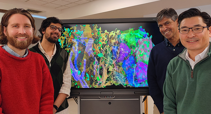
A new study published in Current Biology reveals the nanostructure of brain cells at an unprecedented level of resolution
Brain cells are among the most anatomically complex cells in the human body. They create an intricate web of connections that enables the brain to detect, process, encode and respond to diverse information. Importantly, communication breakdown between brain cells leads to disorders and diseases such as dementia and Alzheimer’s disease that affects an estimated 50 million people worldwide. Until now, our understanding of the elaborate architecture of brain cells and their connectivity has been hampered by the availability of imaging technologies and computational approaches. Without detailed three-dimensional (3D) models incorporating the precise shape and connections of brain cells, researchers cannot fully understand how the brain is built and how it may change following injury or disease.
In a new study published today in Current Biology, researchers from the Research Institute of the McGill University Health Centre (RI-MUHC) and McGill University report the development of new powerful computer vision and machine learning approaches to decode the complex structural framework of brain cells and their intricate three-dimensional physical properties.

Titled “Organizing principles of astrocytic nanoarchitecture in the mouse cerebral cortex,” the study was co-led by Keith Murai, PhD, senior scientist and leader of the Brain Repair and Integrative Neuroscience Program at the RI-MUHC, and Kaleem Siddiqi, PhD, professor in the School of Computer Science and the Centre for Intelligent Machines, McGill University. Bringing together a team of neuroscientists, anatomists, mathematicians and computer scientists, the Murai and Siddiqi labs produced and analyzed some of the largest and highest resolution 3D images of brain cells ever made.
In collaboration with Hojatollah Vali, PhD, and Craig Mandato, PhD, at the Facility for Electron Microscopy Research in the Department of Anatomy and Cell Biology, McGill University, the study used an ultra-high-resolution imaging approach called focused ion beam scanning electron microscopy (FIB-SEM). This yielded thousands of sequential nanometer-thick slices of brain tissue (comparatively, a grain of sand is about one million nanometers in diameter), resulting in detailed cellular reconstructions. With the quantity of raw data acquired, it took the team nearly four years to build 3D models of brain cells.
“This project is a perfect example of the importance of fundamental research, and how it leads to new knowledge that fuels biomedical discoveries to impact human health.”
— Keith Murai, PhD
“We initially wanted to get a clear picture of the complexity of brain cells at a new level of resolution using the FIB-SEM technology available at McGill,” says Chris Salmon, a postdoctoral fellow supervised by Keith Murai. “However, as the project progressed and 3D models of cells developed, we realized the power of the datasets on hand, leading to more complex questions about how the brain is organized.”
The group focused on understanding the fundamental properties of a particular brain cell type called an astrocyte. These cells are likely the most structurally complex cells of the human brain and play a critical role in maintaining brain health, controlling the function of brain circuits, and responding to brain injury and disease.
“At the beginning of the project, there was no expertise available for this kind of high-resolution nanostructural analysis at the RI-MUHC, McGill, or really in Canada,” says Kaleem Siddiqi, PhD, co-leader of the study. “We knew that this would be a long-term, potentially risky project requiring tremendous financial resources and dedication from team members, especially the joint first authors, Chris Salmon, Tabish Syed and Benny Kacerovsky. The computational approaches we developed and used over the course of the study were created completely from scratch. At each step, we had to develop state-of-the-art methods and find the right tools to answer the questions asked.”
Murai adds, “So far, we have only analyzed smaller pieces of brain. However, evolving imaging technologies will soon allow us to analyze larger and more complex areas of the brain. This analysis will harness the cutting-edge approaches that we have developed in this study. Combining computer vision methods with machine learning and artificial intelligence approaches will be key to speeding up the process of resolving the intricate pattern of connections in the brain.”
The Murai-Siddiqi team is nearing completion of another study looking at brain pathology in a model of Alzheimer’s disease. Making the data and analyses part of Open Science and accessible to the global scientific community is also a priority. Murai adds, “This project is a perfect example of the importance of fundamental research, and how it leads to new knowledge that fuels biomedical discoveries to impact human health.”
About the study
Scientific Publication
Christopher K. Salmon, Tabish A. Syed, J. Benjamin Kacerovsky, Nensi Alivodej, Alexandra L. Schober, Tyler F.W. Sloan, Michael T. Pratte, Michael P. Rosen, Miranda Green, Adario Chirgwin-Dasgupta, Shaurya Mehta, Affan Jilani, Yanan Wang, Hojatollah Vali, Craig A. Mandato, Kaleem Siddiqi, Keith K. Murai,
Organizing principles of astrocytic nanoarchitecture in the mouse cerebral cortex,
Current Biology, 2023, ISSN 0960-9822,
https://doi.org/10.1016/j.cub.2023.01.043.
(https://www.sciencedirect.com/science/article/pii/S0960982223000775)
Read the publication in Current Biology.
The authors gratefully acknowledge technical support from the Molecular Imaging Platform at the RI-MUHC and funding support from the Canadian Institutes of Health Research (CIHR), Natural Sciences and Engineering Research Council of Canada (NSERC), Fonds de recherche du Québec – Santé (FRQS), International Development Research Centre (IDRC), Azrieli Foundation, and the Montreal General Hospital Foundation.


